


Cardiac Mapping Systems & 3D Electroanatomical Mapping: How Technology Helps Doctors Navigate the Heart's Electrical Landscape
Cardiac mapping systems — particularly 3D electroanatomical mapping — allow cardiologists to create detailed, colour-coded, three-dimensional models of the heart's electrical activity during catheter-based procedures. By simultaneously recording the position of the catheter and the electrical signals at each point of contact, these systems help pinpoint the precise source of arrhythmias such as atrial fibrillation, atrial flutter, and ventricular tachycardia. The technology enables more targeted ablation, reduces reliance on X-ray imaging, and improves procedural navigation. Its use depends on the complexity of the arrhythmia and the cardiologist's assessment. Always consult a qualified cardiac electrophysiologist for guidance specific to your situation.
At A Glance
Inside every heartbeat is an electrical signal travelling a precise route. When that route goes wrong, arrhythmias follow. Cardiac mapping systems — particularly 3D electroanatomical mapping — give cardiologists a real-time, three-dimensional view of the heart's electrical activity, allowing them to locate the exact source of rhythm problems before and during treatment. This article explains what these systems are, how they work in general terms, who benefits from them, and what patients can expect when this technology is part of their care.
When the Heart's Electrical Map Goes Off-Course
Picture a city's power grid. Every neighbourhood receives electricity through a carefully planned network of cables and substations. Now imagine one rogue cable sending current down a path it was never meant to travel — and every time it does, the entire grid flickers.
That is, in simplified terms, what happens during certain heart rhythm disorders. The heart's electrical system has a precise route it follows with every beat. When abnormal pathways develop — or normal pathways malfunction — the result is an arrhythmia. The challenge for cardiologists has always been finding exactly where the problem originates.
For decades, that search was like navigating a city with no map. Today, 3D electroanatomical mapping systems give physicians something close to a GPS.
What Is Cardiac Mapping?
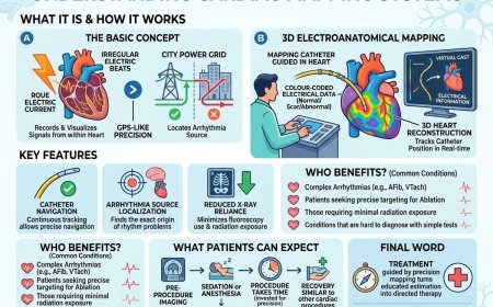
The Basic Concept
Cardiac mapping is the process of recording and visualising electrical signals from within the heart to identify the origin and pathway of arrhythmias. Rather than looking at the heart from the outside, mapping allows cardiologists to see — in real time — where electrical activity is normal, where it is abnormal, and precisely where a treatment such as ablation needs to be delivered.
Mapping can be performed in several ways, ranging from simple surface recordings to sophisticated catheter-based systems. The most advanced of these are three-dimensional electroanatomical mapping platforms, which combine electrical data with anatomical imaging to create a detailed, colour-coded, navigable model of the heart.
Why It Matters
Without accurate mapping, ablation procedures — which destroy abnormal tissue to eliminate faulty electrical pathways — would rely heavily on fluoroscopy (X-ray imaging) alone, offering no information about the electrical characteristics of what is being targeted. Mapping adds a critical second layer of information: not just where the catheter is, but what the heart is doing electrically at that precise location.
How 3D Electroanatomical Mapping Works
Building the Map
During a cardiac catheterisation procedure, one or more mapping catheters are guided into the heart through blood vessels. As the catheter moves across the inner surface of the heart, it simultaneously records two types of data:
- Electrical signals — the timing, voltage, and pattern of electrical activity at each point contacted
- Positional data — the precise three-dimensional location of the catheter tip within the heart
These two streams of data are combined by the mapping system's software to construct a three-dimensional model of the heart chamber being studied. The model is colour-coded to show areas of normal electrical activity, abnormal activity, scarred tissue, and the pathways along which electrical signals are travelling.
What the Cardiologist Sees
The result is a dynamic, rotatable, three-dimensional representation of the heart — almost like a virtual cast of the chamber — overlaid with electrical information. Cardiologists can zoom in, rotate, and analyse the map to identify the precise origin of an arrhythmia and plan where treatment should be delivered.
Some advanced systems can also integrate pre-procedural imaging — such as CT or MRI scans of the heart — into the map, giving the physician an even more detailed anatomical reference.
Tracking the Catheter in Real Time
One of the most clinically valuable features of electroanatomical mapping is catheter navigation. The system continuously tracks the position of the catheter within the heart and displays it on the map in real time. This allows the cardiologist to navigate with precision, return accurately to specific sites, and confirm that the ablation catheter is in the right location before delivering treatment — all with significantly reduced reliance on X-ray exposure.
Who Benefits from 3D Electroanatomical Mapping?
Mapping systems are typically used when:
- The arrhythmia is complex or has multiple sources
- Standard surface ECG or simpler recordings have not been sufficient to localise the problem
- An ablation procedure is planned and precise targeting is essential
- The arrhythmia involves a large area of abnormal tissue, such as in some cases of ventricular tachycardia or persistent atrial fibrillation
- Minimising radiation exposure during the procedure is a priority
Conditions for which 3D mapping is commonly employed include atrial fibrillation, atrial flutter, ventricular tachycardia, and complex supraventricular arrhythmias. Whether mapping is used in your specific case — and which system is used — depends on your cardiologist's assessment and the resources of the treating centre.
What to Expect as a Patient
If your cardiologist recommends a procedure that includes electroanatomical mapping, you will not experience the mapping process separately from your overall procedure. From a patient's perspective, the experience of a mapping-guided ablation is broadly similar to any catheter-based cardiac procedure:
- You will be sedated or under general anaesthesia
- Catheters will be inserted through blood vessels, typically in the groin
- The procedure takes place in a specialised cardiac catheterisation or electrophysiology laboratory
- The total duration is longer than procedures performed without mapping, as building the map takes time — but that time is invested in greater precision
- Recovery is generally similar to other catheter-based cardiac procedures, with a short hospital stay
The mapping system itself is operated by a team of electrophysiology specialists and cardiac technologists who work alongside your cardiologist throughout the procedure.
Questions to Ask Your Cardiologist
- Will 3D mapping be used during my procedure, and why or why not?
- What type of arrhythmia do I have, and how does mapping help identify its source?
- How does the information from mapping change how my treatment is planned or delivered?
- How long is the procedure expected to take when mapping is included?
- What does the team's experience with mapping-guided procedures look like?
- Are there any additional risks associated with the mapping process itself?
Common Misconceptions
"Cardiac mapping is its own separate procedure I have to undergo." In most cases, mapping is performed as part of the same procedure as the treatment itself — typically ablation. Patients do not generally undergo a separate mapping session in advance.
"The map shows a picture of my heart like an X-ray." Electroanatomical maps are not anatomical images in the traditional sense. They are data visualisations — colour-coded models that represent electrical information overlaid on a reconstructed three-dimensional shape. They are tools for the cardiologist, not photographs of the heart.
"If my arrhythmia requires mapping, it must be very serious." The use of mapping reflects the complexity of the arrhythmia and the precision required for treatment — not necessarily the severity of the condition. Many patients who undergo mapping-guided procedures have manageable conditions that simply require a targeted approach.
"Mapping guarantees the ablation will work." Mapping significantly enhances precision, but it does not guarantee a specific outcome. Results depend on the nature of the arrhythmia, the extent of abnormal tissue, and individual patient factors. Your cardiologist will discuss realistic expectations.
Professional Support
If your doctor has recommended a cardiac procedure involving mapping technology, or you want to understand whether an electrophysiology evaluation may be right for your situation, our specialists can help you ask the right questions and navigate your options.
👉 https://myamericandoctor.com/our-doctors/
You may also choose to enroll in our upcoming concierge medical clinic in India, Global Concierge Doctors. We offer U.S.-style primary care with 24/7 access to India-based physicians. When required, we coordinate referrals to trusted cardiac specialists in India and the U.S.
The Final Word
The heart is a four-chambered electrical organ that beats over 100,000 times a day. When its wiring goes wrong, the consequences can range from uncomfortable to life-altering. The development of 3D electroanatomical mapping represents one of the most significant advances in how cardiologists find and fix those problems — turning what was once a process of educated estimation into one of guided precision.
If an ablation procedure has been recommended for you, understanding that your cardiologist may be working with one of the most sophisticated navigation tools in modern medicine is worth knowing. The next step is a conversation with a specialist who can apply that technology to your individual situation.
Reader Poll
What brings you to this article?
- I've been diagnosed with an arrhythmia and want to understand my treatment options
- My cardiologist has recommended an ablation procedure and I'm doing my research
- I'm curious about how cardiac technology works
- I'm researching on behalf of a family member or patient
If this article helped you, share it with someone navigating a cardiac diagnosis.
Medical Disclaimer
For Informational Purposes Only
This article is intended for general educational and informational purposes only. It does not constitute medical advice, diagnosis, or treatment. References to cardiac mapping systems, 3D electroanatomical mapping, catheter ablation, electrophysiology studies, CT or MRI cardiac imaging integration, or any related cardiac procedure are for informational purposes only and do not represent a recommendation for any specific diagnostic test, procedure, or treatment approach.
Not a Recommendation for Specific Tests or Treatments
All decisions regarding cardiac investigation and treatment — including whether mapping-guided ablation is appropriate, which mapping platform is used, and how care is structured — must be made by a qualified cardiologist or cardiac electrophysiologist in consultation with the individual patient, based on their complete medical history, clinical findings, and individual circumstances.
Seek Professional Medical Advice
Always seek the advice of a qualified physician or other licensed healthcare provider with any questions regarding a medical condition, diagnostic procedure, or treatment. Do not disregard professional medical advice or delay seeking it based on information in this article.
Emergency Situations
If you are experiencing chest pain, severe palpitations, loss of consciousness, fainting, or any other cardiac emergency, call your local emergency services immediately.
AI-Assisted Content Disclosure
This article was developed with the assistance of AI-based writing tools under the editorial direction and review process of MyAmericanDoctor.com. Content is reviewed for general accuracy and alignment with established educational standards.
Category: Cardiology | Suggested Subcategory: Cardiac Electrophysiology & Interventional Cardiology
Sources Consulted
- American Heart Association (AHA) — Catheter ablation and electrophysiology studies. www.heart.org
- Heart Rhythm Society (HRS) — Patient resources on electrophysiology and cardiac mapping. www.hrsonline.org
- National Heart, Lung, and Blood Institute (NHLBI) — Cardiac catheterisation and arrhythmia treatment overview. www.nhlbi.nih.gov
- Mayo Clinic — Cardiac ablation: Tests and procedures. www.mayoclinic.org
- Cleveland Clinic — Electrophysiology study (EPS) and cardiac mapping. my.clevelandclinic.org
What's Your Reaction?
 Like
0
Like
0
 Dislike
0
Dislike
0
 Love
0
Love
0
 Funny
0
Funny
0
 Angry
0
Angry
0
 Sad
0
Sad
0
 Wow
0
Wow
0